Microcirculation in the Brain
(The real purpose of the scientific method is to make sure nature hasn’t misled you into thinking you know something you actually don’t know.*)
Overview
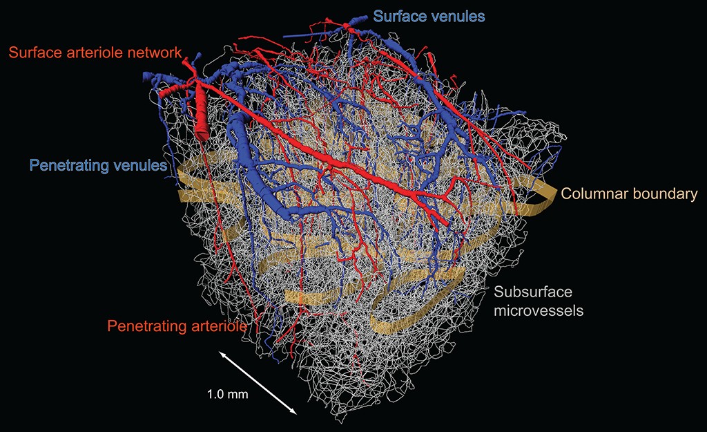
Neuronal activity is intimately linked with changes in metabolism and subsequent changes in regional blood flow. Even minor interruptions in flow can adversely effect cognitive function. We have completed the connectome of brain vascualture to understand structral determinates of brain perfusion.
We continuing to investigate the modulation of brain perfusion by rhythmic vasodynamics along with the coupling of neuronal signaling to arterioles.


Ongoing Projects
Ongoing projects address: (i) The role of vaso-oscillations in patterning perfusion of the brain; (ii) The role of brainstem area RVLM in modulating cortical perfusion. (iii) The nature of blood recovery by the venuole system. (iv) Single neuron energetics with respect to signaling and glucose, lactate, and oxygen uptake. (v) Extension of lessons from the rodent on vaso-oscillations to human fMRI (w/Polimeni laboratory)
Confluence of tools
The combination of many in vivo tools, including electrophysiology, intracellular ion measurements, neurotransmitter receptor activation measurements, microdialysis measurements, and single vessel blood flow measurements, allow us to assess the influence of single neurons and networks of neurons on vascular control. These experiments are further supported by optical activation and pharmacological manipulations to derive causal relations, and our automated reconstruction techniques to map local architectonics.